

On This Day in Health: November 12, 1881
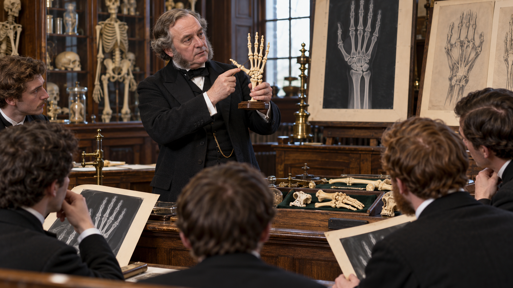
On November 12, 1881, Dublin surgeon Edward Hallaran Bennett presented specimens of an intra-articular fracture at the base of the first metacarpal, later called the Bennett fracture. This date matters because it fixes a broad medical story to a specific moment, place, and group of people. It also shows how health history is shaped carefully by institutions and practical choices as well as laboratory discoveries.
The background was already developing before that day. Thumb injuries were easy to dismiss as simple sprains or routine fractures, yet the saddle-shaped carpometacarpal joint carries major forces during pinch and grasp. Without imaging, subtle displacement and joint instability were difficult to recognize. Those conditions defined what participants could attempt and what patients or communities could reasonably expect. The available tools, social expectations, and professional structures all shaped the result. Change also depended on cooperation among people whose roles were not always recorded equally. They explain why later accounts sometimes compress years of preparation into a single anniversary. Looking at that preparation keeps credit broad and makes the turning point easier to understand without turning it into a miracle story.
What happened on the date was consequential but limited. Bennett used anatomical specimens and clinical observation to show a characteristic fracture fragment with displacement of the metacarpal by muscle pull. The description connected joint mechanics with a predictable deforming pattern. An eponym does not describe every thumb-base fracture, and later work distinguished comminuted Rolando patterns and other injuries. Bennett's nineteenth-century patients lacked modern radiographs, fixation, anesthesia, hand therapy, and outcome measures. The distinction is important: the event did not instantly solve the underlying health problem, and later progress depended on follow-up research, training, policy, funding, and public trust. Contemporary evidence also helps separate what participants knew then from interpretations added after the result became famous.
Its longer legacy reaches beyond the original setting. Recognition of the unstable pattern helped guide reduction and immobilization and later supported surgical fixation when alignment cannot be maintained. Current care uses dedicated imaging, assessment of joint congruity and stability, shared decisions about operative or nonoperative treatment, and rehabilitation aimed at motion, strength, and return to daily use. Seen in context, the anniversary is useful because it connects a concrete historical episode with continuing questions about evidence, access, safety, and responsibility in health care. That connection is stronger when the people affected, the limits of the achievement, and the work still unfinished remain part of the account.
Bennett used anatomical specimens and clinical observation to show a characteristic fracture fragment with displacement of the metacarpal by muscle pull. The description connected joint mechanics with a predictable deforming pattern.
An eponym does not describe every thumb-base fracture, and later work distinguished comminuted Rolando patterns and other injuries. Bennett's nineteenth-century patients lacked modern radiographs, fixation, anesthesia, hand therapy, and outcome measures.
Recognition of the unstable pattern helped guide reduction and immobilization and later supported surgical fixation when alignment cannot be maintained.
Thumb injuries were easy to dismiss as simple sprains or routine fractures, yet the saddle-shaped carpometacarpal joint carries major forces during pinch and grasp. Without imaging, subtle displacement and joint instability were difficult to recognize.
Dublin surgeon Edward Hallaran Bennett presented specimens of an intra-articular fracture at the base of the first metacarpal, later called the Bennett fracture.
Current care uses dedicated imaging, assessment of joint congruity and stability, shared decisions about operative or nonoperative treatment, and rehabilitation aimed at motion, strength, and return to daily use.
Explore more of "On This Day ..."
Discover more events from the same date across news, politics, technology, sports, and other fields. Each link highlights significant moments that shaped history on different fronts.