

Colonoscopy Explainer
Colonoscopy is an examination of the inside of the rectum and colon using a long, flexible instrument with a light and camera. It lets a clinician inspect the bowel lining directly, rather than infer its appearance from symptoms alone. The procedure can investigate problems such as unexplained bleeding or persistent changes in bowel habits. It can also be used for colorectal cancer screening. Its purpose is determined by the clinical question, so two people having the same examination may be following quite different care pathways.
The camera sends images to a monitor as the clinician guides the scope through the large intestine. Small tools passed through the instrument can take tissue samples or remove certain polyps. A polyp is a growth on the bowel lining; finding one does not automatically mean cancer. Laboratory examination helps identify what was removed. This combination of direct viewing and sampling is an important distinction from tests that can show a possible abnormality but cannot collect tissue during the same appointment.
Bowel preparation is part of the examination itself. Remaining stool can hide the lining and make findings harder to interpret. The clinical team provides specific instructions about food, fluids, prescribed preparation, and medicines. These instructions should be followed as supplied rather than replaced with an online schedule. Sedation is commonly used, and recovery arrangements depend on what was given. Planning transport and understanding the discharge instructions helps make the procedure a complete episode of care instead of an isolated test.
Colonoscopy has limitations and risks, including bleeding, a tear in the bowel wall, and reactions to sedation. A clear view improves the examination but does not guarantee that every abnormality will be found. Immediate observations and laboratory results may arrive at different times. The next screening or surveillance interval depends on the findings, examination quality, and medical history. Understanding what the test can answer, what remains uncertain, and who will explain the results helps patients participate in follow-up without treating a single report as the entire story.
Screening examines people without symptoms, while a diagnostic colonoscopy investigates a particular concern. Surveillance means checking again after a previous finding or because of an established risk. These terms describe why the test is being done, not different kinds of camera. Knowing which pathway applies makes a discussion about the examination more useful and helps explain the follow-up plan.
Other colorectal tests have different strengths. Stool testing looks for particular signals in a sample, while CT colonography constructs images using X-rays. A positive result may require a conventional colonoscopy. An alternative test is therefore not always the end of the process. The relevant question is which sequence of tests can answer the clinical concern with acceptable burdens and risks.
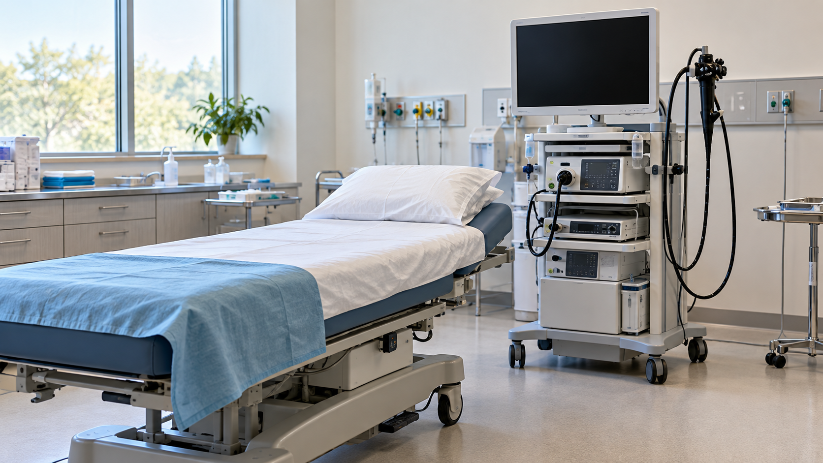
The examination usually takes place in a hospital or outpatient service. Staff check the medical history, medicines, consent, and preparation before the scope is introduced. The patient is positioned on an examination couch, and the bowel is gently expanded for visibility. The clinician examines the lining and documents findings, including whether any samples or polyps were taken.
Preparing for the appointment also means confirming practical details. Patients can ask when they will receive preparation instructions, whom to contact if they cannot complete them, and what assistance is needed afterward. A written medication list helps the team identify relevant issues. These conversations support the prescribed plan; they do not justify independently stopping medicines or improvising a different preparation.
A visual finding and a tissue diagnosis are related but distinct. A clinician may describe a polyp immediately, while a laboratory later reports its microscopic features. Follow-up recommendations can depend on both. Waiting for a tissue result is not evidence that the finding is necessarily serious, but it is a reason to know how and when the final explanation will arrive.
Aftercare instructions identify symptoms that need prompt assessment and explain temporary restrictions following sedation. Severe pain, substantial or persistent bleeding, or other concerning changes should not be dismissed as routine recovery. The examination is most useful when results reach the patient and any additional care is arranged. Good communication matters alongside the technical quality of the procedure.
Explore more "Explainers"
Discover additional explainers across politics, science, business, technology, and other fields. Each explainer breaks down a complex idea into clear, everyday language—helping you better understand how major concepts, systems, and debates shape the world around us.