

MRI Explainer
Magnetic r
esonance imaging, or MRI, creates detailed images of structures inside the body using a strong magnetic field, radiofrequency signals, and a computer. It does not use X-rays or ionizing radiation. MRI is useful for many soft tissues, including the brain, spinal cord, joints, and organs. The examination is chosen to answer a clinical question; its detail does not make it the best test for every possible problem.
The scanner interacts with hydrogen nuclei, which are abundant in the water and other molecules of body tissues. A magnetic field influences their alignment, and radiofrequency pulses temporarily alter it. As they return toward their earlier state, signals are detected and used to construct images. Different tissue properties and scanning sequences produce different kinds of contrast, allowing the same body area to be viewed in several ways.
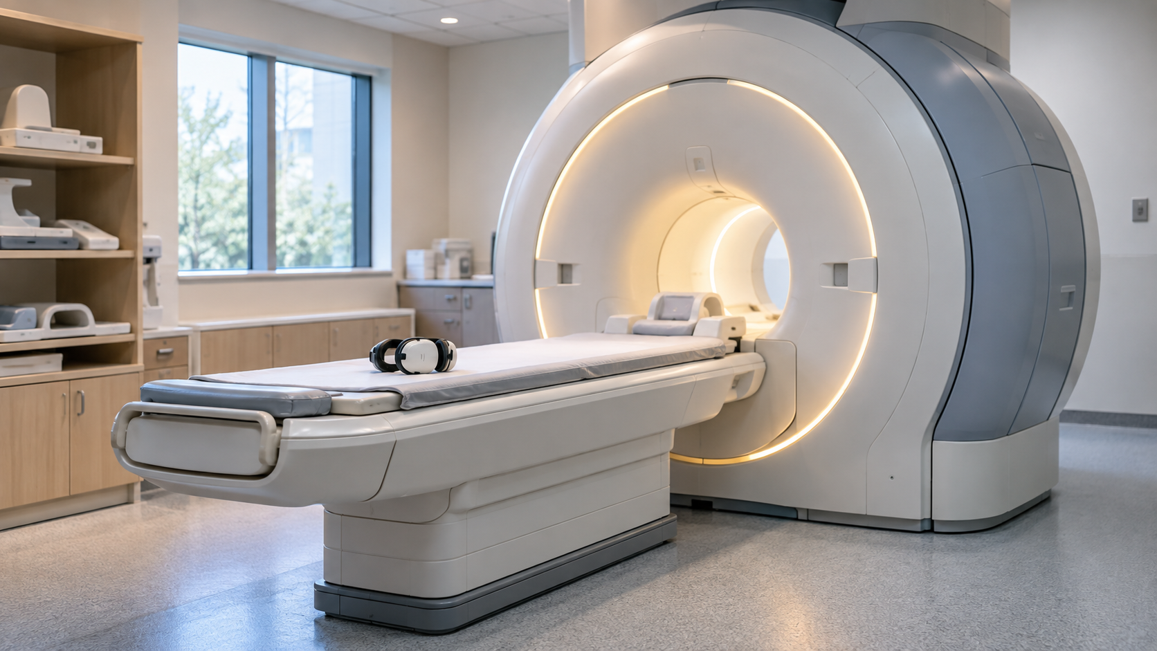
An examination usually involves lying on a table that moves into the scanner while a series of images is collected. Remaining still helps avoid blurred images. The machine can make loud knocking sounds, so hearing protection is important. The team can communicate with the patient during the scan. Some people find the enclosed space uncomfortable; discussing anxiety beforehand helps the team plan appropriate support.
Safety screening is essential because the strong magnet can interact with objects and implanted devices. Patients should tell the team about implants, previous procedures, metal fragments, and other relevant circumstances. Some scans use an injected contrast agent to help answer the question, while others do not. A radiologist interprets the images alongside the reason for the examination, and the referring clinician explains how the findings fit the wider assessment.
MRI is not a single photograph taken from outside the body. It gathers signals through a sequence of measurements and reconstructs images. Different sequences emphasize different tissue characteristics, and images may be produced in several planes. A scan protocol is selected for the body area and suspected problem. That is why two MRI appointments can differ in duration or technique.
Detailed imaging can help diagnose disease, investigate symptoms, or monitor a known condition. Nevertheless, more detail does not automatically produce a more useful answer. CT, ultrasound, or another test may be better suited to a particular question. The choice balances the information needed, the person’s circumstances, and practical limitations rather than ranking every imaging method from good to bad.
The magnetic field can exert strong forces on certain metal objects and affect some devices. Screening determines whether an implant is safe for the specific scanner and conditions. Having an implant does not automatically mean MRI is impossible, but the team needs accurate information. A device described as conditional requires its stated conditions to be met, not a casual assumption that all devices are equivalent.
External objects and clothing also matter. Jewelry, electronic devices, and items with metal components must be managed according to the facility’s instructions. The magnetic environment remains important even when images are not being collected. Patients should follow the team’s guidance rather than independently deciding that a familiar object or an earlier scan proves there is no risk this time.
Contrast can make some tissues or abnormalities easier to distinguish. Whether it is useful depends on the question, and its use requires attention to factors such as kidney function and a history of reactions. MRI without contrast and MRI with contrast are not interchangeable in every situation. A person should discuss relevant health information with the team rather than assuming contrast is always necessary or always avoidable.
Movement, the ability to tolerate the examination, and other factors can limit image quality. Findings may also be incidental or require comparison with other tests. The report describes what was seen and the radiologist’s interpretation; it does not replace the clinical history. A useful follow-up explains which findings answer the original question, which are uncertain, and whether any further assessment is needed.
Explore more "Explainers"
Discover additional explainers across politics, science, business, technology, and other fields. Each explainer breaks down a complex idea into clear, everyday language—helping you better understand how major concepts, systems, and debates shape the world around us.