

Retinal Detachment Explainer
Retinal detachment happens when the retina, the light-sensitive tissue at the back of the eye, separates from its normal position. This can threaten permanent vision loss and requires urgent assessment. Sudden flashes, a marked increase in floaters, or a dark curtain or shadow across vision are warning signs. Contact an eye specialist or emergency service right away rather than waiting for a routine appointment or for pain to develop.
The retina helps convert incoming light into signals used for sight. In the most common form of detachment, a tear lets fluid pass behind it and lift it away. Changes in the gel inside the eye as people age can contribute to a tear. Other forms involve scar tissue pulling on the retina or fluid collecting behind it without a break. These mechanisms have different causes, so treatment is guided by examination.
Risk is higher in people with severe nearsightedness, previous eye injury or surgery, and certain retinal disorders. A personal or family history can also matter. Risk factors do not mean detachment is inevitable, and their absence cannot rule it out. Many people have longstanding floaters, but a sudden new pattern deserves prompt attention. A small detachment may initially cause limited symptoms, making an appropriate eye examination especially important.
Treatment may seal a tear or involve surgery to return the retina to position. The choice depends on the type, location, and extent of the problem. Early assessment can help preserve vision, but successful reattachment does not guarantee complete restoration of sight. Recovery and visual outcome vary. Follow-up, activity restrictions, and any positioning instructions from the surgical team are part of treatment, rather than optional additions.
In a tear-related detachment, changes in the vitreous gel can create a pull on the retina. If a break forms, fluid can travel through it. This process explains why a new burst of floaters or flashes can be important, even though those symptoms also occur with other eye changes. A person cannot safely decide from symptoms alone whether a harmless gel change, a retinal tear, or a detachment is responsible.
Tractional detachment occurs when scar tissue draws the retina away from its support. Diabetic retinopathy is one possible setting. Exudative detachment involves fluid accumulation without a retinal tear and can be associated with inflammation or leaking vessels. Knowing the mechanism helps the specialist plan care. The similar-sounding names describe different physical problems, rather than three interchangeable labels for the same procedure or prognosis.
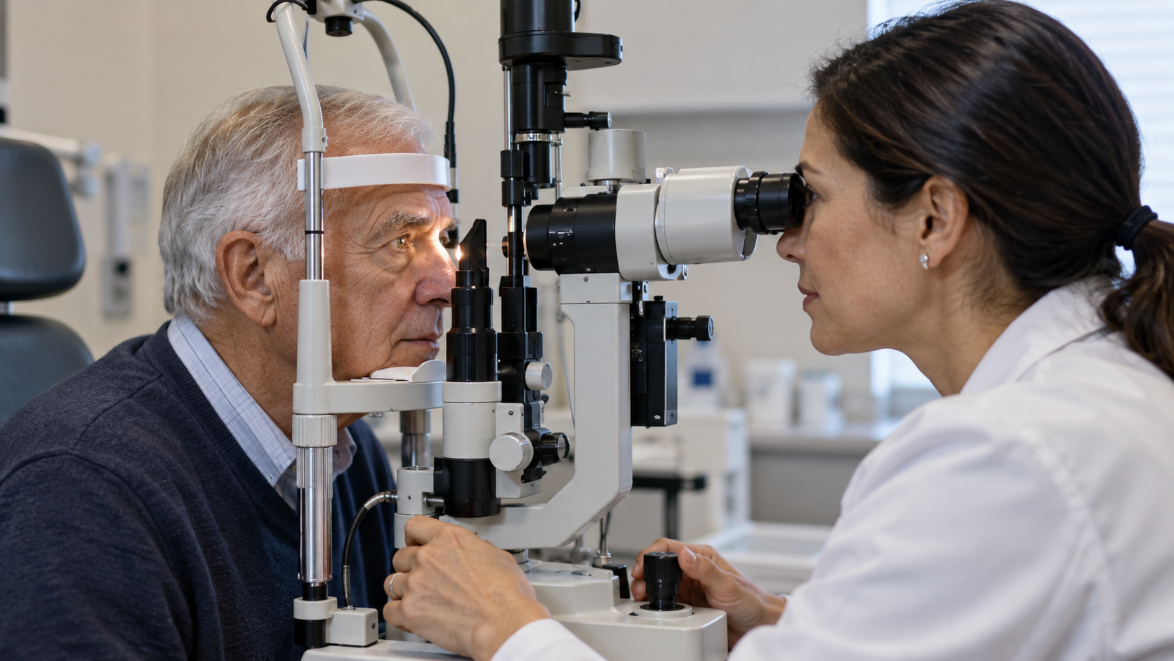
A dilated eye examination widens the pupil so the clinician can inspect the retina. The specialist may need to examine its edges carefully. Ultrasound or optical coherence tomography can provide additional information in selected cases. These investigations help locate and characterize the problem. They are performed by trained professionals; looking at the eye in a mirror or checking a phone photograph cannot establish that the retina remains attached.
If sight changes suddenly, arranging safe transport may be necessary. Continue to seek urgent help even when symptoms fluctuate or no pain is present. Routine appointments for existing eye disease should not delay a new emergency assessment. Protective eyewear can reduce injury risk during hazardous activities, and regular recommended eye examinations support ongoing care, but neither precaution eliminates every possible cause of detachment.
A small retinal tear may be treated with a laser or a freezing procedure to create a seal around it. A detached retina can require pneumatic retinopexy, scleral buckling, vitrectomy, or a combination. These approaches respectively use a bubble, external support, or work on the internal gel to help restore position. The eye surgeon explains which approach fits the findings and whether treatment to seal tears is also needed.
When a gas bubble is used, the team may prescribe a particular head position and restrictions on flying. The exact instructions depend on the operation and must come from the treating team. Vision may remain blurred during recovery, and some people need further surgery. Worsening pain, redness, or vision after treatment should be reported promptly. Returning symptoms also require assessment, because a repaired retina can develop another problem later.
Explore more "Explainers"
Discover additional explainers across politics, science, business, technology, and other fields. Each explainer breaks down a complex idea into clear, everyday language—helping you better understand how major concepts, systems, and debates shape the world around us.