

How CT Scans Build Cross-Sectional Images Explainer
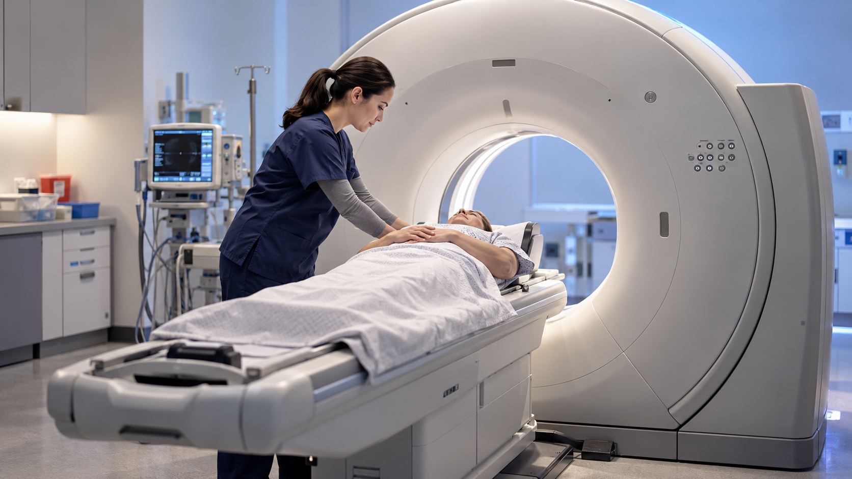
Computed tomography, or CT, makes cross-sectional images using X-rays measured from many directions. A patient lies on a motorized table that moves through the circular opening of a gantry. Inside, an X-ray tube rotates opposite rows of detectors. Each measurement records how much the beam is weakened along one path through the body, a property called attenuation.
Dense materials such as bone absorb or scatter more X-rays than air or many soft tissues. The scanner gathers a large set of projections as the tube turns. A computer uses mathematical reconstruction to estimate the attenuation at each small volume element, or voxel. Display settings then map those values to shades of gray suited to lungs, bone, brain, or other tissues.
Modern scanners commonly use helical acquisition: the table moves continuously while the tube rotates, creating a spiral path of data. Multiple detector rows capture many slices at once. Thin slices can be stacked into three-dimensional views or reformatted in different planes. These displays come from the same measured volume and do not create information that was never captured.
Contrast agents make selected structures easier to distinguish. Iodine in blood vessels or organs absorbs X-rays strongly, while oral contrast may outline the digestive tract. The choice depends on the clinical question and patient risks. Timing matters because contrast moves through circulation, so scans may be synchronized to emphasize arteries, veins, or particular organs.
CT uses ionizing radiation, so protocols balance image quality against dose. Tube current, voltage, scan length, pitch, body size, and reconstruction methods all affect exposure and noise. Motion, metal, incomplete data, and very dense material can create artifacts that mimic or hide anatomy. Technologists and radiologists recognize and reduce these limitations.
Unlike a single projection X-ray, CT separates overlapping structures into slices, making internal location and shape clearer. It can be fast enough for trauma, stroke, lung, and vascular imaging. The result is not a direct photograph; it is a calibrated mathematical estimate based on X-ray transmission, instrument geometry, and reconstruction choices.
A rotating X-ray source and detector array measure attenuation through the body from many angles.
Reconstruction estimates attenuation in voxels and displays selected ranges as grayscale slices.
Helical acquisition and multirow detectors rapidly collect a three-dimensional volume.
Contrast agents and carefully timed scans highlight vessels, organs, or the digestive tract.
Protocols manage radiation dose while noise, motion, metal, and dense material can cause artifacts.
CT images are computed estimates that separate anatomy hidden by overlap in ordinary X-rays.
Explore more "Explainers"
Discover additional explainers across politics, science, business, technology, and other fields. Each explainer breaks down a complex idea into clear, everyday language—helping you better understand how major concepts, systems, and debates shape the world around us.