

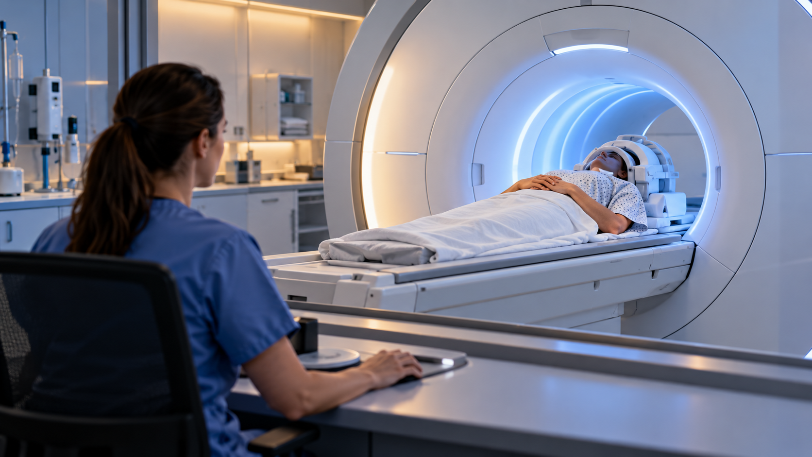
How MRI Builds Images of the Body Explainer
Magnetic resonance imaging relies heavily on hydrogen because the body contains abundant water and fat. Hydrogen nuclei behave like tiny magnetic moments. In the scanner’s strong static magnetic field, slightly more of them align with the field than against it, producing a small net magnetization. The nuclei also precess at a frequency determined by field strength.
A radiofrequency coil sends a pulse tuned near that resonant frequency. The pulse tips the net magnetization away from the main field and brings many nuclei into a coordinated phase. When the pulse stops, the system moves back toward equilibrium. The changing magnetic signal induces a voltage in receiver coils placed near the body.
Two relaxation processes shape the signal. T1 relaxation describes recovery along the main field, while T2 relaxation describes loss of phase coherence across the transverse plane. Tissues differ in their T1 and T2 behavior and in proton density. By adjusting pulse timing, an MRI sequence can emphasize particular contrasts among fat, fluid, muscle, brain tissue, or pathology.
Gradient coils add small, controlled variations to the magnetic field across three directions. Those gradients make resonant frequency and phase depend on location. Repeating measurements with different gradient patterns encodes spatial information into the signal. Mathematical reconstruction, commonly based on Fourier methods, converts the recorded data into slices or three-dimensional volumes.
Special sequences can measure flowing blood, water diffusion, chemical composition, or changes associated with neural activity. Contrast agents may alter local relaxation and make selected structures more visible. The image is therefore not a direct photograph; it is a map built from magnetic signals under a defined sequence, and its appearance depends on acquisition choices.
MRI does not use ionizing X-rays, but the magnet and radiofrequency energy require strict safety procedures. Ferromagnetic objects can become dangerous projectiles, some implants need special evaluation, and loud gradient switching requires hearing protection. Motion can blur data, scans can be lengthy, and interpretation depends on trained clinicians and the broader medical context.
The main magnet creates a small net alignment of hydrogen nuclei, especially those in water and fat.
A radiofrequency pulse tips and synchronizes the nuclei, and receiver coils detect their returning signal.
T1 and T2 relaxation differ among tissues and supply much of the image contrast.
Magnetic field gradients encode position in frequency and phase so a computer can reconstruct anatomy.
Special sequences map flow, diffusion, chemistry, or functional changes rather than only static structure.
MRI avoids ionizing radiation but still requires screening for metal, implants, heating, noise, and motion.
Explore more "Explainers"
Discover additional explainers across politics, science, business, technology, and other fields. Each explainer breaks down a complex idea into clear, everyday language—helping you better understand how major concepts, systems, and debates shape the world around us.