

How Ultrasound Builds Images Inside the Body Explainer
Diagnostic ultrasound uses sound waves above human hearing to image structures inside the body without ionizing radiation. A transducer contains piezoelectric material that converts electrical pulses into mechanical vibrations and returning vibrations into electrical signals. The probe repeatedly sends short pulses and listens for echoes, allowing many measurements to be assembled into a moving image in real time.
Sound travels through tissue until it encounters a boundary where acoustic impedance changes. Some energy is reflected and some continues. The scanner estimates depth from the time between transmission and echo, using an assumed average sound speed, while echo strength influences brightness. Repeated lines at different angles create a two-dimensional slice; arrays can steer and focus the beam electronically.
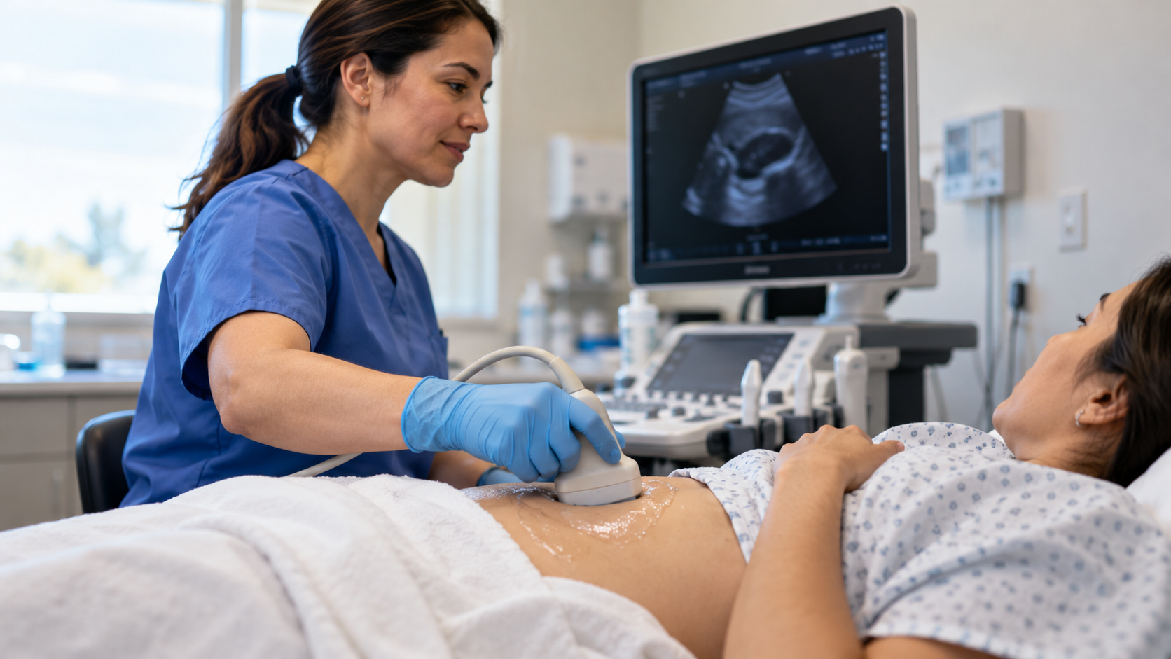
Coupling gel removes air between the probe and skin because air reflects most ultrasound at that boundary. Frequency creates a tradeoff: higher frequencies have shorter wavelengths and can reveal finer detail but are absorbed more quickly, while lower frequencies penetrate deeper with less spatial resolution. The sonographer selects probe, angle, focus, gain, and depth for the target.
Doppler ultrasound uses frequency shifts from moving red blood cells to estimate the direction and speed of flow. Color and spectral displays summarize motion but depend on beam angle, sampling, and processing assumptions. Other modes track tissue motion or stiffness. These are measured signals rendered by software, not direct color photographs of anatomy or blood.
Bone and gas block or scatter sound, limiting views of the adult brain, lungs, and structures behind bowel gas. Reflection, refraction, attenuation, multiple echoes, and incorrect assumed speed can create artifacts that mimic or obscure anatomy. Some artifacts are useful clues, but interpretation requires training and correlation with other views and clinical information.
Diagnostic ultrasound has a strong safety record, yet sound deposits energy and can cause slight heating or mechanical effects. Good practice uses the lowest output and shortest exposure that provides needed information, especially in pregnancy. Ultrasound matters because a compact transducer can turn echo physics into real-time anatomical and functional information at the bedside, in clinics, and during procedures.
Piezoelectric elements convert electrical pulses to sound and returning echoes to signals.
Echo time estimates depth while echo strength helps determine image brightness.
Gel removes an air gap that would otherwise reflect most sound.
Higher frequency improves detail but reduces penetration depth.
Doppler shifts reveal motion and flow but depend strongly on beam angle.
Gas, bone, attenuation, and artifacts limit interpretation despite the lack of ionizing radiation.
Explore more "Explainers"
Discover additional explainers across politics, science, business, technology, and other fields. Each explainer breaks down a complex idea into clear, everyday language—helping you better understand how major concepts, systems, and debates shape the world around us.