

How Electron Microscopes Reveal Tiny Structures Explainer
Electron microscopes use accelerated electrons instead of visible light to probe a specimen. Electrons have wave-like behavior, and at typical operating energies their wavelength is far shorter than visible light, permitting much finer spatial detail in principle. Resolution still depends on lens aberrations, beam brightness, stability, detector performance, sample preparation, scattering, and radiation damage rather than wavelength alone.
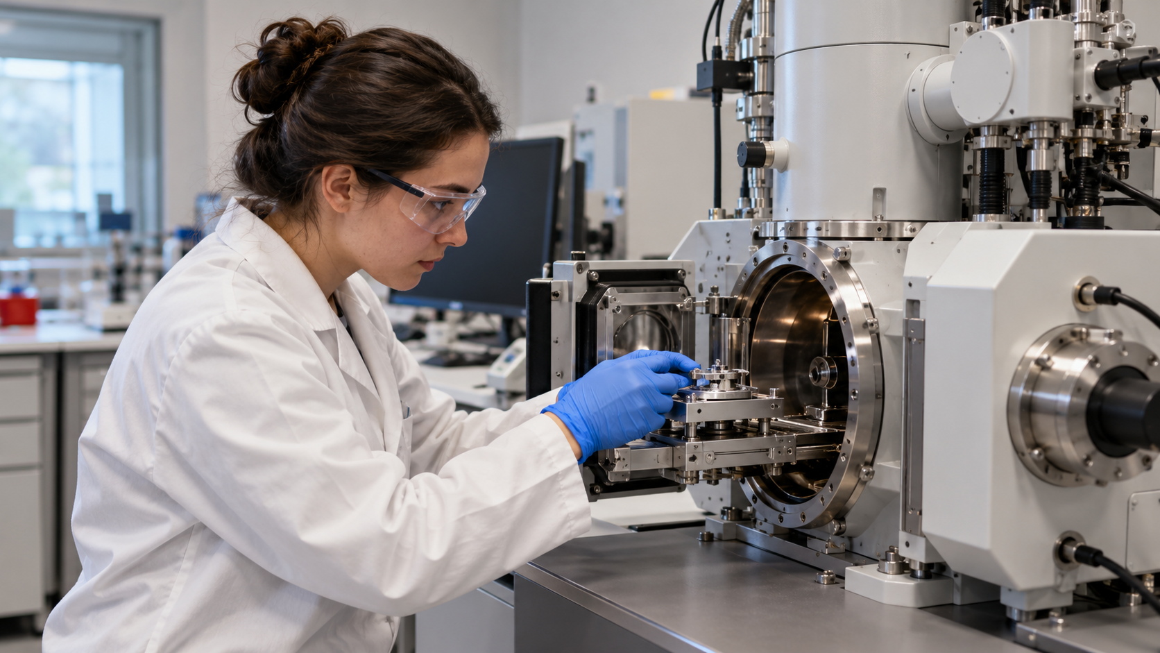
An electron source emits a beam that electromagnetic lenses and apertures shape and focus inside a high vacuum. Vacuum prevents electrons from scattering from air molecules and protects the source. Unlike a glass lens bending light, magnetic and electric fields steer charged electrons. Mechanical vibration, stray fields, contamination, and thermal drift can blur measurements at very small scales.
In a scanning electron microscope, a focused beam moves point by point over a surface. Beam interactions release low-energy secondary electrons that are sensitive to surface shape and higher-energy backscattered electrons influenced by composition. Detectors convert changing signal intensity into a raster image. X-rays generated by the beam can identify elements, but the displayed image is not a conventional photograph.
In a transmission electron microscope, electrons pass through an extremely thin specimen. Scattering and phase changes carry information about internal structure, crystal arrangement, and composition. Lenses form images or diffraction patterns, and scanning transmission modes focus a small probe across the sample. Preparing a representative section thin enough for transmission can be one of the hardest steps.
Biological samples may be fixed, dehydrated, stained with heavy atoms, frozen rapidly, or reconstructed from many low-dose images. Nonconductive SEM samples may need a thin conductive coating or low-vacuum methods. Every preparation can alter structure, and electron beams can heat, charge, displace atoms, or break chemical bonds. Apparent texture and contrast must therefore be interpreted with method controls.
Electron microscopy can reveal viruses, organelles, catalysts, defects, crystal lattices, and semiconductor features, often with elemental or spectroscopic information. It does not automatically show living activity or true color. The instrument creates a measurement from electron-specimen interactions, and reliable conclusions depend on calibration, multiple views, careful preparation, dose control, and comparison with complementary techniques.
Short electron wavelengths permit finer potential resolution than visible light.
Vacuum, electromagnetic lenses, apertures, and stable stages control the beam.
SEM scans surfaces and measures secondary, backscattered, or X-ray signals.
TEM transmits electrons through thin specimens to reveal internal and crystal structure.
Preparation and electron dose can alter, charge, heat, or damage the sample.
Electron images are signal maps requiring calibration and interpretation, not ordinary color photographs.
Explore more "Explainers"
Discover additional explainers across politics, science, business, technology, and other fields. Each explainer breaks down a complex idea into clear, everyday language—helping you better understand how major concepts, systems, and debates shape the world around us.