

Mammography Explainer
Ma
mmography uses low-dose X-rays to produce images of breast tissue. It can be used for screening before symptoms appear or to investigate a lump, another breast change, or an earlier imaging finding. Those are different clinical purposes, even when the same machine is used. A mammogram can reveal changes that deserve closer attention, but an image alone does not always establish whether a finding is cancer.
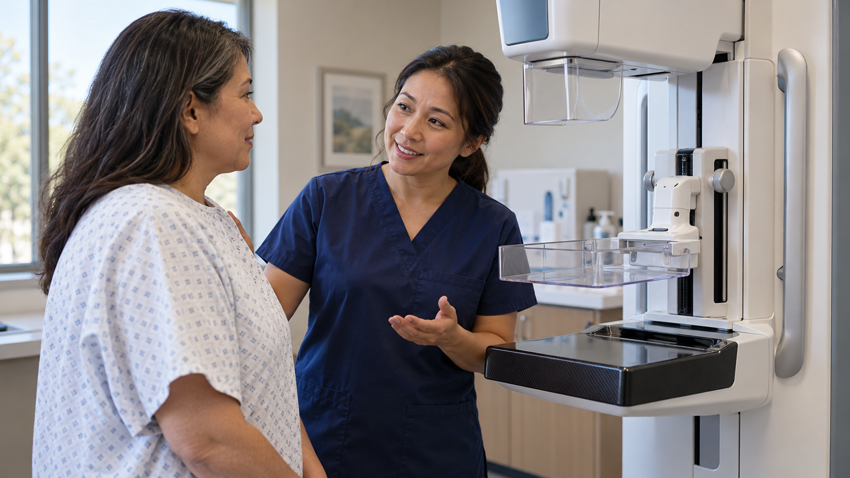
During the examination, the breast is positioned on a support and compressed with a paddle while images are taken. Compression spreads out overlapping tissue, helps keep it still, and improves image quality. It can be uncomfortable, so communication with the technologist matters. The breast is repositioned for different views. Digital breast tomosynthesis, sometimes called 3D mammography, collects images from several angles and reconstructs them into thin views through the tissue.
A radiologist examines the images for findings such as masses, calcifications, or changes compared with previous examinations. The report also describes breast density, meaning the relative amounts of fibrous and glandular tissue compared with fat. Dense tissue can make cancers harder to see on mammograms and is associated with increased breast cancer risk. Density is an imaging characteristic, not something that can be determined by how a breast feels.
Results guide the next step. Some findings need no additional investigation, while others call for more imaging, follow-up, or a biopsy. A callback is not the same as a cancer diagnosis, and many callbacks do not lead to one. Screening plans depend on age, individual risk, and applicable guidance. A new breast change should be discussed with a clinician even after a recent reassuring screening result.
Screening looks for possible disease in people without breast symptoms. Diagnostic mammography addresses a specific question, such as a new lump or an area requiring a closer look after screening. It may involve additional views and other imaging. The distinction helps explain why two people can have different appointments or follow-up plans without receiving inconsistent care.
Preparation instructions come from the imaging facility. Products on the breast or underarm area can interfere with interpretation, so facilities commonly provide guidance about them. Previous images can be valuable for comparison, and the team should know about implants or prior breast procedures. Telling the technologist about discomfort helps the examination proceed as effectively and comfortably as possible.
Dense and fatty tissues appear differently on a mammogram. Dense tissue can obscure a finding, which is one reason a normal mammogram cannot guarantee that no cancer exists. Density also contributes to risk assessment. Additional imaging may be considered in some circumstances, but the decision depends on the full risk picture and the benefits and limitations of the additional test.
Mammography has both benefits and limitations. Screening can find cancers earlier, but can also produce false alarms, miss a cancer, or identify a cancer that would not have caused harm during a person’s lifetime. These possibilities help explain why screening recommendations involve tradeoffs. The best plan is not determined solely by having the newest imaging technology or the greatest number of examinations.
Reports often use BI-RADS, a standardized system that connects an imaging assessment with a recommended next step. An incomplete assessment can mean additional images are needed. A suspicious finding may lead to biopsy, which examines tissue rather than its appearance alone. The category is a communication tool for the care team, not a cancer stage or a substitute for pathology.
Following the recommended next step matters more than trying to interpret one unfamiliar word in isolation. A clinician can explain the finding, how it compares with earlier images, and what should happen next. Symptoms and imaging are considered together. If results or follow-up instructions do not arrive as expected, contacting the facility helps prevent an unresolved examination from being mistaken for a completed assessment.
Explore more "Explainers"
Discover additional explainers across politics, science, business, technology, and other fields. Each explainer breaks down a complex idea into clear, everyday language—helping you better understand how major concepts, systems, and debates shape the world around us.