

On This Day in Health: July 29, 2010
On July 29, 2010, The Lancet published online a proof-of-concept study showing that a damaged articular joint surface could regenerate in rabbits by recruiting the animals' own cells. A team led by Columbia University researcher Jeremy Mao designed a biodegradable scaffold shaped like the rounded end of a rabbit's upper arm bone. Instead of growing replacement tissue from transplanted stem cells in a laboratory, the researchers used the scaffold as a temporary structure and added a signaling protein to attract cells already present in the body. The result suggested a different route for tissue engineering: guide repair where the injury exists.
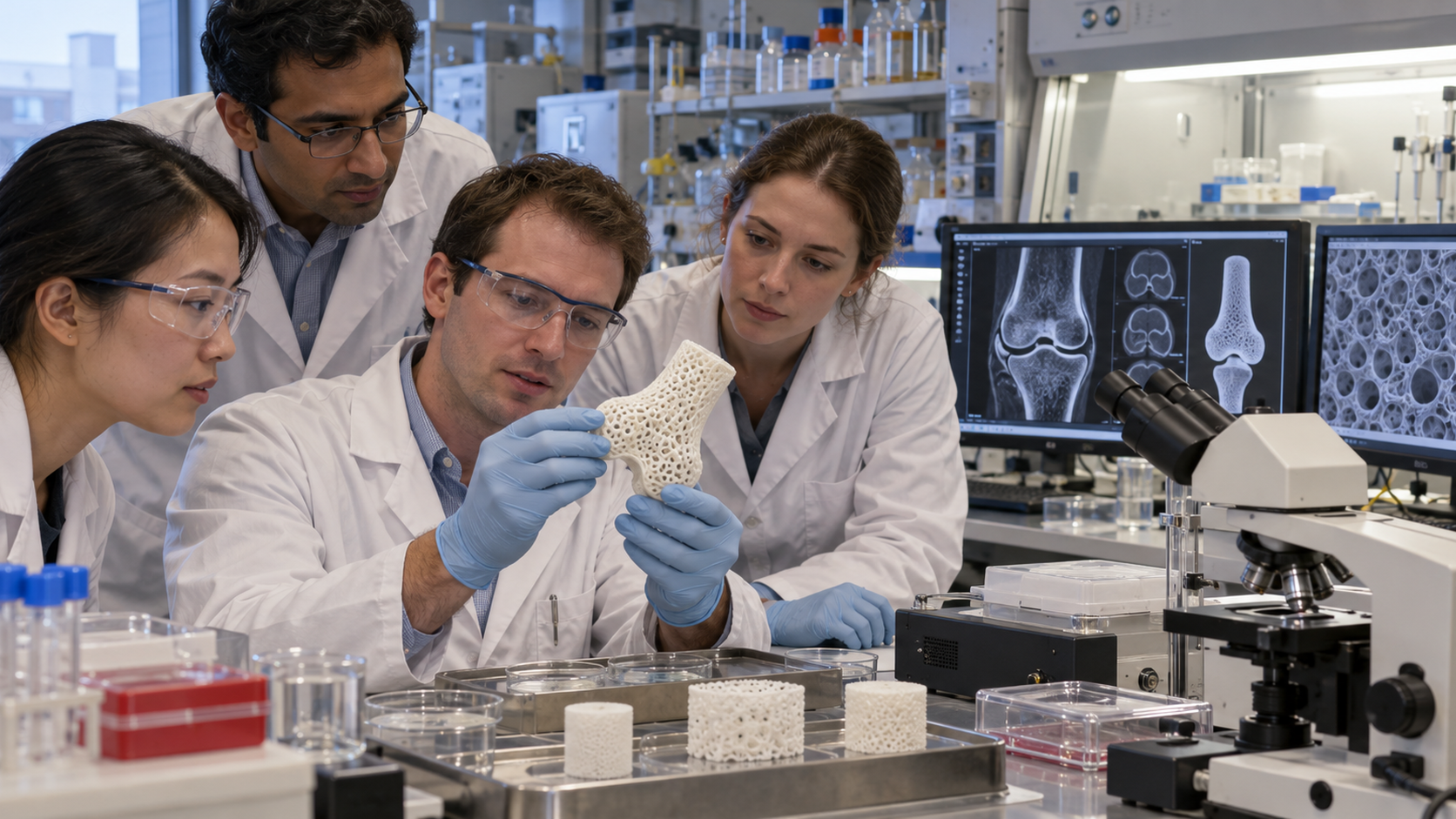
The investigators first captured the joint's shape with laser scanning and computer-aided design. They fabricated a porous scaffold from polycaprolactone and hydroxyapatite, materials selected to support tissue growth and gradually break down. Some scaffolds received collagen gel containing transforming growth factor beta 3, or TGF-beta 3, a protein involved in cartilage development. Others received gel without the factor, while a small comparison group had the joint surface removed without receiving a scaffold. The experiment tested whether anatomical structure and a localized biological cue could draw endogenous cells into a complex site and organize both cartilage and underlying bone.
Rabbits receiving TGF-beta 3 scaffolds resumed weight bearing and movement within three to four weeks more consistently than animals in the other groups. After four months, the treated surfaces showed regenerated cartilage integrated with regenerated bone. The growth-factor group produced thicker, denser cartilage and recruited substantially more cells than scaffolds without the protein. Its measured compression and shear properties approached those of normal cartilage. The untreated defects did poorly. These results were striking because smooth, load-bearing cartilage has little natural capacity to heal, and an entire joint surface combines tissues with different structures, blood supplies, and mechanical demands.
The study did not create a ready treatment for human arthritis. It involved rabbits, limited observation, surgically controlled injuries, and a scaffold designed for one anatomical site. Researchers still needed to test durability, safety, scaling, immune responses, and performance under human loads. Even so, the report offered evidence that cell homing might complement or sometimes avoid cell transplantation. It helped shift regenerative medicine toward instructive materials that shape the body's response rather than merely replacing damaged parts. July 29 marks a careful experimental milestone: a functioning articular surface regenerated in an animal model, with promise defined by the need for much more research.
The team used laser scanning and computer-aided design to make a porous scaffold matching the rabbit humeral joint surface that researchers had removed.
A collagen gel containing TGF-beta 3 supplied a local biological signal intended to recruit the animal's own cells and encourage cartilage formation.
Rabbits with growth-factor scaffolds regained movement more consistently, and four-month samples showed cartilage integrated with regenerated underlying bone.
The experiment was small and preclinical; it did not establish safety, durability, manufacturing methods, or effectiveness for people with osteoarthritis or traumatic injuries.
The work supported cell homing as a regenerative strategy in which implanted materials guide endogenous cells instead of delivering laboratory-expanded stem cells.
Later research has continued to examine scaffold chemistry, growth signals, mechanics, and patient-specific geometry while confronting the difficulty of durable human cartilage repair.
Explore more of "On This Day ..."
Discover more events from the same date across news, politics, technology, sports, and other fields. Each link highlights significant moments that shaped history on different fronts.