

On This Day in Health: August 28, 1919
Godfrey Newbold Hounsfield was born near Newark, Nottinghamshire, England, on August 28, 1919. He joined the Royal Air Force during the Second World War, worked with radar and electronics, and later became an engineer at EMI’s Central Research Laboratories. Hounsfield was interested in using computers to reconstruct information that could not be seen directly. In the late 1960s, he proposed measuring how X-rays passed through an object from many angles and using calculations to produce a cross-sectional image rather than the overlapping shadows of a conventional radiograph.
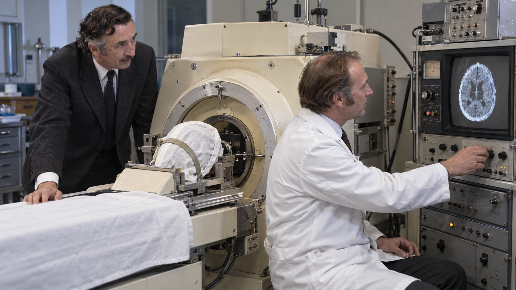
The concept required sensitive detectors, stable X-ray sources, extensive computation, and a practical scanning geometry. Hounsfield built experimental systems and worked with neuroradiologist James Ambrose at Atkinson Morley’s Hospital in London. The first clinical brain scan was performed in 1971 on a patient with a suspected tumor. The image took hours to acquire and reconstruct by standards now considered extraordinarily slow, but it distinguished soft tissues inside the skull with clarity unavailable from ordinary X-rays. A new diagnostic field emerged rapidly.
South African physicist Allan Cormack had independently developed mathematical theory for reconstructing cross-sections from transmission measurements. Hounsfield and Cormack shared the 1979 Nobel Prize in Physiology or Medicine for the development of computer-assisted tomography. CT scanners soon became faster, expanded from the head to the whole body, and improved through rotating detector arrays and better algorithms. The Hounsfield unit, a scale of X-ray attenuation used in CT images, bears his name. Modern scans can guide emergency care, cancer staging, surgery, biopsy, and treatment planning.
CT also exposes patients to ionizing radiation, and contrast agents can carry risks, so appropriate use matters. Clinicians balance diagnostic benefit against dose, age, pregnancy, kidney function, allergies, and alternatives such as ultrasound or MRI. Equipment and specialist access also remain unequal. Hounsfield’s birthday marks a medical advance created by engineering, mathematics, computing, and clinical collaboration. His central idea turned many imperfect measurements into an anatomical slice, changing diagnosis without an incision. The continuing obligation is to use that power carefully: choose the right scan, minimize dose while preserving needed information, and ensure that images improve decisions rather than simply adding tests. CT’s speed has changed emergency medicine especially. A scan completed in minutes can reveal internal bleeding, stroke patterns, fractures, or organ injury while teams prepare treatment. That speed can save time, but incidental findings may also trigger anxiety and more testing. Good imaging practice includes explaining what a scan can answer and how results will change care.
Hounsfield was born on August 28, 1919, developed expertise in radar and electronics, and later worked in computing at EMI research laboratories.
He proposed collecting X-ray transmission measurements from many angles and using a computer to reconstruct a cross-sectional image.
Collaboration with neuroradiologist James Ambrose produced the first clinical brain scan in 1971 and revealed soft-tissue detail unavailable from standard radiographs.
Allan Cormack’s independent mathematical work supplied complementary foundations, and the two shared the 1979 medicine prize for computed tomography.
CT now supports emergency diagnosis, cancer care, image-guided procedures, surgical planning, and quantitative assessment across the body.
Because CT uses ionizing radiation and sometimes contrast agents, safe practice requires appropriate indications, optimized dose, and patient-specific risk assessment.
Explore more of "On This Day ..."
Discover more events from the same date across news, politics, technology, sports, and other fields. Each link highlights significant moments that shaped history on different fronts.