

CT Scans Explainer
Computed tomography, or CT, uses X-rays and computer processing to create cross-sectional pictures of the body. A conventional X-ray combines structures into a flat projection; CT collects information from many angles and reconstructs a series of slices. Clinicians can examine those slices or combine them into other views. This helps reveal the location and extent of abnormalities that are difficult to separate on a single projection. The scan answers a particular clinical question rather than providing an unlimited assessment of health.
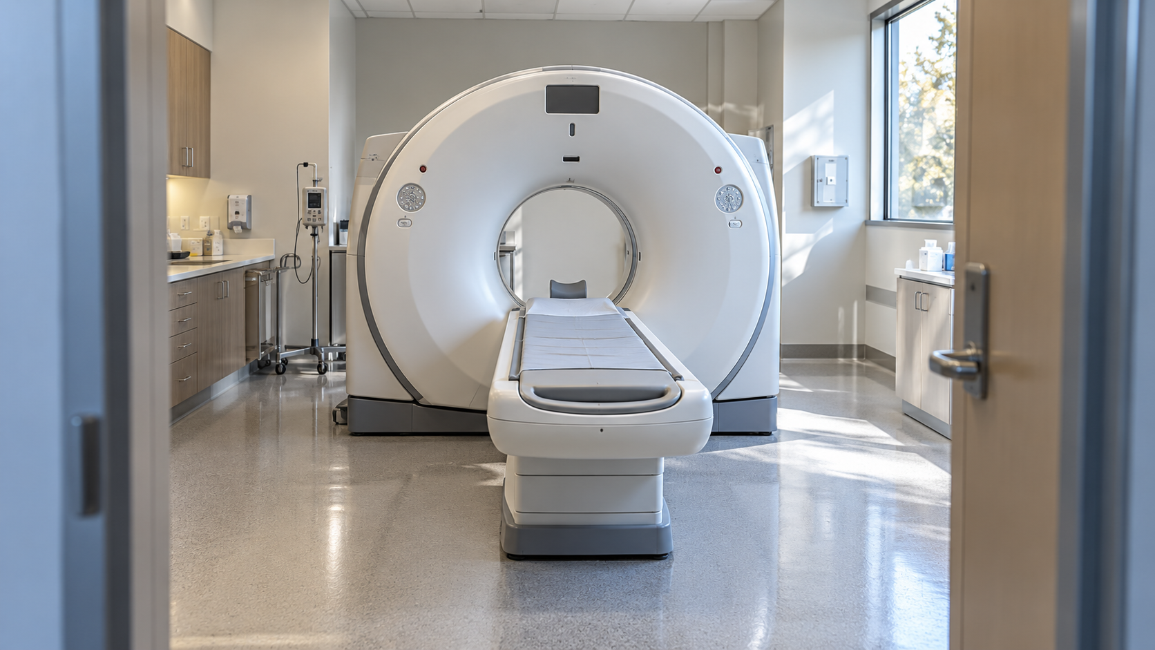
During a scan, a motorized table moves through a short circular opening while the X-ray source and detectors collect measurements around the body. Remaining still helps produce clear images. Some examinations use contrast material, which can make blood vessels or other structures easier to distinguish. Contrast may be given in different ways depending on the examination. It is not required for every CT scan, and the decision considers the information needed together with relevant medical history and possible risks.
CT is useful in many settings, including evaluating certain injuries, investigating symptoms, planning treatment, and monitoring a known condition. It can acquire images quickly, but speed does not make it the best test for every concern. Ultrasound, MRI, or another approach may offer a more suitable answer in a particular situation. CT and MRI also use different physical methods: CT involves ionizing radiation, while MRI uses magnetic fields and radio waves. The appropriate choice depends on the tissue and question being examined.
The main tradeoff is obtaining useful diagnostic information while avoiding unnecessary exposure and procedures. Radiation dose depends on the examination and equipment settings, and contrast introduces separate considerations. A radiologist interprets the images in context, including symptoms and earlier studies when available. An unexpected finding may need clarification without necessarily representing dangerous disease. Knowing why the scan was ordered, whether preparation is required, and who will explain the report helps keep a detailed image connected to meaningful clinical decisions.
The term tomography refers to imaging in sections. CT uses measured differences in how tissues absorb X-rays to reconstruct those sections. The resulting pictures are representations built from data, not ordinary camera photographs taken inside the body. Understanding this helps explain why image quality, movement, and reconstruction methods can affect what a scan shows.
A modern CT scanner usually has a wide, short gantry rather than the long enclosed tunnel associated with some MRI systems. The table and scanner work together to collect the required area. A scan of one body region does not automatically assess everything else. The protocol is tailored to the question so that useful information can be obtained efficiently.
Before scanning, staff may ask about pregnancy, previous contrast reactions, kidney problems, and medicines. Which details are relevant depends on the planned examination. Some scans have preparation instructions and others do not. The patient should follow the imaging service’s guidance rather than assume that a previous scan’s preparation applies to a new appointment with a different protocol.
A radiologist reviews the images and produces a report describing findings and their possible significance. The clinician who requested the examination connects that report with the rest of the medical assessment. A phrase such as indeterminate means additional context may be needed. It is neither a definite diagnosis nor a reason to ignore recommended follow-up.
Ionizing radiation can increase longer-term risk, which is weighed against the expected benefit of the information. Children and repeated examinations may require particular care in choosing protocols. Useful imaging is not made safer by being avoided when urgently needed, but unnecessary imaging adds burden without equivalent benefit. The decision is about clinical value, not simply acquiring more pictures.
Whole-body scanning offered without a specific indication can produce incidental findings and additional testing. A broad image does not reliably settle every health concern. Questions about what the scan can change in care, whether another method could answer the question, and how results will be communicated support an informed discussion. The goal remains appropriate information for a meaningful next step.
Explore more "Explainers"
Discover additional explainers across politics, science, business, technology, and other fields. Each explainer breaks down a complex idea into clear, everyday language—helping you better understand how major concepts, systems, and debates shape the world around us.