

X-rays Explainer
An X-ray is a form of electromagnetic radiation used to produce images of structures inside the body. A detector records the pattern after the beam passes through the area being examined. Different tissues absorb different amounts, creating an image that a clinician can interpret. Dense bone usually appears lighter, while air appears darker. The result is useful medical information, but it is not a photograph that reveals every kind of tissue problem equally well. The test must match the clinical question.
Conventional X-rays are commonly used to assess suspected fractures and other bone problems. They can also help examine teeth, lungs or certain abdominal concerns. The term X-ray covers a wider family of techniques: CT combines multiple projections into cross-sectional images, and fluoroscopy can display movement during a procedure. Those examinations differ in purpose and radiation exposure. Being told that a test uses X-rays does not, by itself, explain what the appointment involves or how its risk compares with another examination.
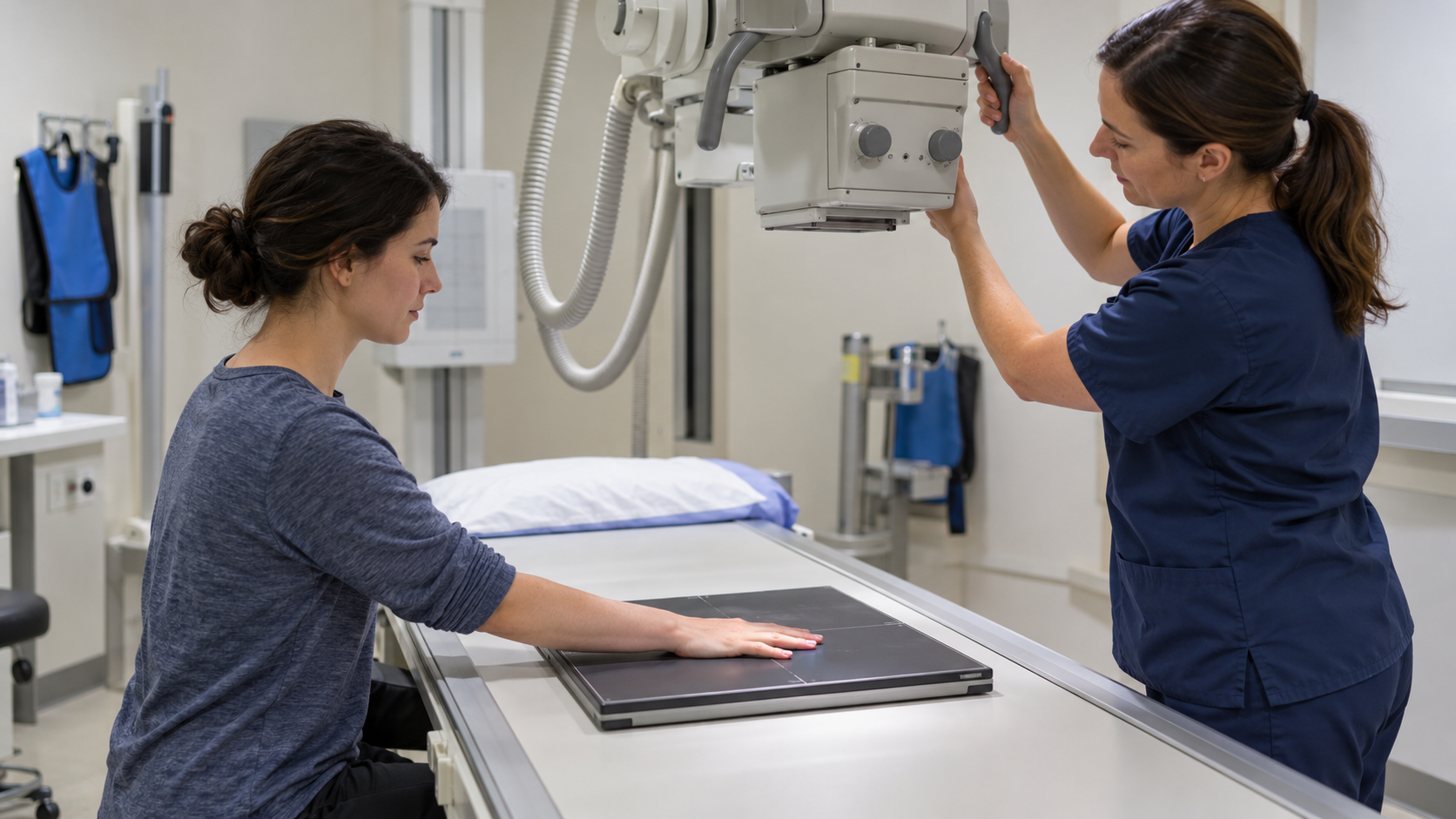
For a straightforward examination, a technologist positions the relevant part of the body near the detector and aligns the equipment. You may need to hold still, briefly hold your breath or move into another position for a different view. The actual exposure is quick and cannot be felt. An injured limb may be uncomfortable to position, so tell staff about pain or mobility limitations. Following their instructions helps produce a clear image while avoiding an unnecessary repeat caused by movement.
Medical X-rays use ionizing radiation, which carries a small potential increase in future cancer risk. The dose depends on the examination, and children are more sensitive to radiation than adults. A medically appropriate test usually offers a benefit that outweighs the small risk. The aim is to answer a useful clinical question with an appropriate technique and dose. Ask why the image is needed and tell the team about possible pregnancy and relevant previous imaging, rather than refusing a necessary test without discussing the circumstances.
Preparation depends on the examination. A simple bone X-ray usually needs little preparation, although jewellery or metal near the area may need to be removed. Other tests may involve fasting or contrast material and have different instructions. Read the appointment information and ask if anything is unclear. Do not apply preparation rules from a previous scan to a different procedure without checking.
Tell the team if you are pregnant or might be pregnant, and mention medicines, allergies and any previous reaction to contrast material. These details help staff plan the examination appropriately. They do not automatically mean imaging must be cancelled. When contrast is part of the procedure, the team should explain what it is for and whether additional preparation or observation is required.
X-rays are particularly useful for showing bone, but provide less detail about many muscles, tendons and ligaments. Some subtle fractures may not appear on the initial image. Depending on symptoms and examination findings, a clinician may recommend another view, follow-up imaging or a different technique. A normal image should be interpreted with the injury history rather than treated as a universal guarantee that nothing is wrong.
A radiologist reviews the images and sends a report to the referring clinician, who can explain the findings and next steps. Ask when and how results will be provided. The image may support a diagnosis, guide treatment or indicate that another test is needed. If symptoms persist or worsen, contact the care team even when the initial report did not identify a clear explanation.
Radiation exposure is kept as low as reasonably achievable while preserving enough image quality to answer the question. Appropriate settings depend partly on body size and the area examined. Checking previous imaging can sometimes avoid an unnecessary duplicate, but repeat imaging may also be justified when circumstances change. Useful questions concern the expected benefit, suitable alternatives and whether the result will influence care.
Ultrasound and MRI do not use ionizing radiation, yet they are not interchangeable replacements for every X-ray examination. The best choice depends on what clinicians need to see, the urgency and the patient's circumstances. Broad comparisons of machine names can be misleading without that context. An informed discussion connects the particular test with its purpose and explains both its limits and the practical next step after the result.
Explore more "Explainers"
Discover additional explainers across politics, science, business, technology, and other fields. Each explainer breaks down a complex idea into clear, everyday language—helping you better understand how major concepts, systems, and debates shape the world around us.